Immunohistochemistry (IHC) for Oncology
我们的临yaboapp体育官网床前肿瘤学团队还提供内部组织学和免疫组化(IHC)除了其他in vitro services for oncology,进一步扩展我们广泛的服务清单,以增强您的学习。通过简单地将样品在我们最先进的设施中向组织学实验室移动到组织学实验室来确保样品的最佳保存和样品的结构完整性。
Our high-quality IHC assays give you a time-efficient read-out on quantitative and qualitative data and can be produced expeditiously that reveal the following:
- Efficacy of your compound in target tissues
- 生物标志物在组织中的位置
- Incidence of tumor metastasis
- Occurrence of tissue transformation into tumors
- 肿瘤组织的起源
- 公关ognosis of the animals in the study
特色IHC文章:
Services:
- Normal Tissue
- 肿瘤组织
- Xenograft
- 新标记验证
- 免疫荧光
- 免疫细胞化学
- 多路复用 - 定制标记,最多四种免疫荧光
验证了IHC标记:

AE1 / AE3 + 5d3细胞质阳性小鼠皮肤。细胞角蛋白是上皮细胞中可检测到的丝状蛋白质。它们可用于肿瘤分化,细胞键入和微转型。

CD3.membrane staining in spleen

4T1中的CD3染色
A20中的CD3染色
CT26中的CD3染色
Mouse CD3 membrane staining in spleen. CD3 is used to identify T cells in normal tissue, as well as T cell neoplasms.
CD4.staining in subcutaneous A20
A20中的CD4免疫荧光染色

CT26中的CD4染色
Pan02中的CD4染色
Mouse CD4 membrane staining in mouse tumor tissue. CD4 is used to identify T helper cells, and plays a role in T cell activation.

在皮下CT26小鼠肿瘤组织中染色小鼠CD8膜染色。CD8用于鉴定细胞毒性T细胞。

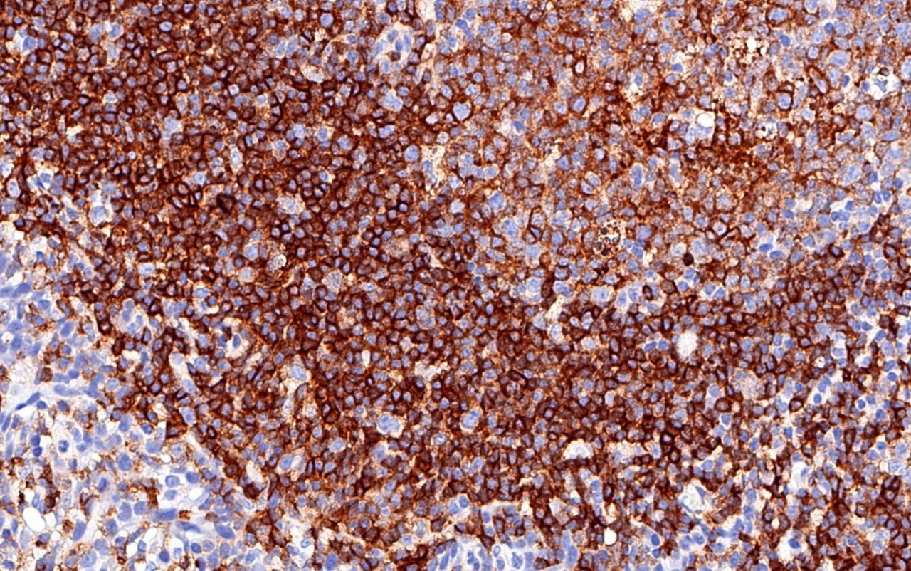
Human CD19 membrane staining in human tonsil. CD19 is a biomarker for B cells, and plays a role in B cell development.

人cd20染色在人的扁桃体。CD20是一种B细胞标记,其功能是实现最佳的B细胞免疫应答。
Human CD22 staining in human tonsil. CD22 is found on the surface of mature B cells that functions as an inhibitory receptor for B cell receptor signaling. It is an active target for B cell malignancies and autoimmune disease.
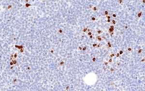
CD38染色4T1

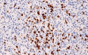
CD3.8 staining in A20
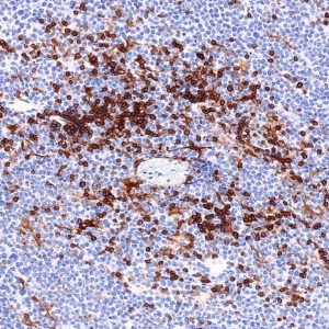
CT26中的CD38染色
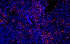
CD3.8 staining in ID8
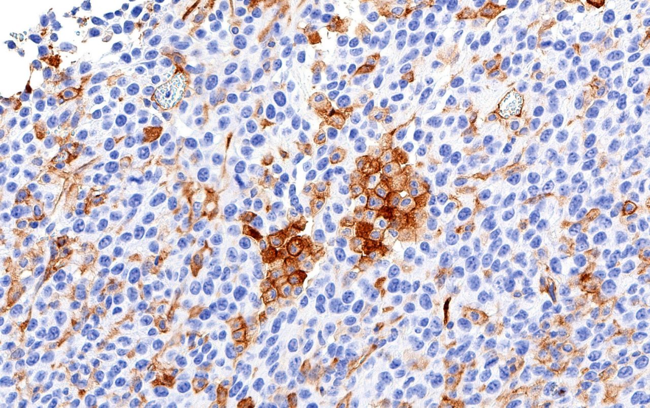
如图所示,在多个组织中染色的小鼠CD38染色。CD38在许多免疫细胞表面上发现(CD4+and CD8+T cells, B cells, and natural killer cells). It has functions in cell adhesion, signal transduction and calcium signaling and is used as a prognostic marker in leukemia patients with CLL.

小鼠CD45膜染色在A20肿瘤组织中。CD45在调节T-和B细胞抗原受体信号传导中起作用。
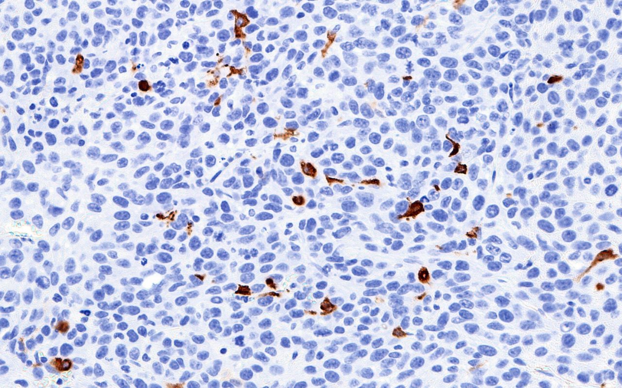
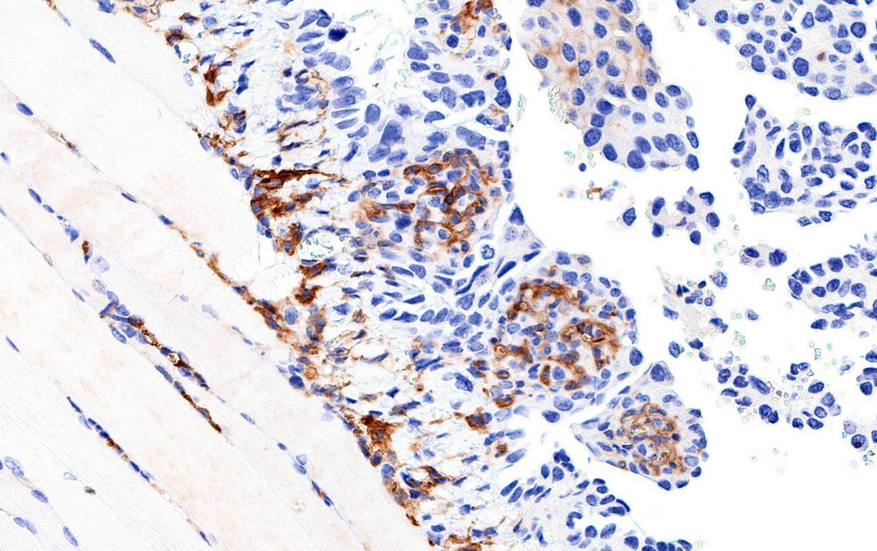
CT26中的CD163染色
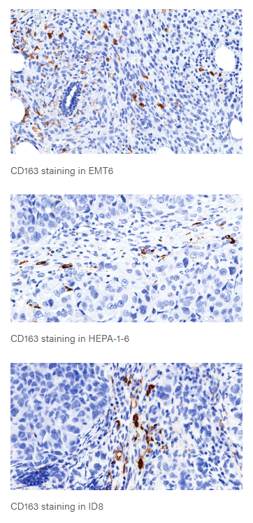
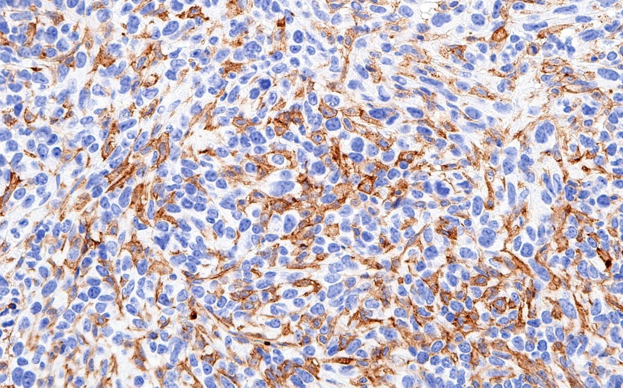
Mouse CD163 staining in multiple tissues, as indicated. CD163 is expressed in macrophages and monocytes. Expression may be indicative of inflammation.

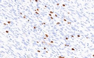
Mouse FOXP3 nuclear staining in spleen. FoxP3 is involved in the development and inhibitory function of regulatory T cells.
Her2 positive cytoplasm in BT474 tumor tissue.

HER2.免疫荧光of SK-OV-3 Cells
HER2是人们参与激酶介导的信号传导途径的激活的人EGF受体。它被用作癌症治疗治疗中的预测因子和决定因素。

人体扁桃体的人类Ki-67核染色。KI-67蛋白质与细胞增殖相关,可以用作癌症中的预后工具。
人类公关积极核BT474肿瘤组织。公关ogesterone receptor is involved in activating signaling pathway for hormone stimulation. It can be utilized in determining appropriate drug therapy in cancer treatment.
