流式细胞仪使用新型CompdC™面板综合树突细胞分析
作者:
David Draper,PHD |科学发展助理主任
日期:
2019年2月
T细胞介导的抗肿瘤反应铰链铰接呈现肿瘤相关抗原(TAA)的抗原呈递细胞(APC)的活性,并为T细胞提供共刺激信号。这反过来激活肿瘤特异性T细胞,触发它们的扩张和招募在肿瘤中,在他们发挥其效果。树突细胞(DC)是在此过程中有用的专业APC。可以激活DC以表达许多性刺激配体,可以将抗原内化并将免疫肿瘤肽均为CD4+和CD8.+T细胞。The development of novel therapeutics that harness DC function is an area of intensive investigation. To facilitate these efforts and address the growing need for robust comprehensive DC immunophenotyping in murine pre-clinical models, Covance has configured a new standard panel, CompDC™. In this Tech Spotlight, we will demonstrate how various DC subsets in tumor and other tissue-derived cell samples can be analyzed using this panel.
For a general introduction to the different DC subsets and their function in cancer biology, read our recent blog“使用同工免疫肿瘤模型进行树突细胞生物学和分析的介绍”
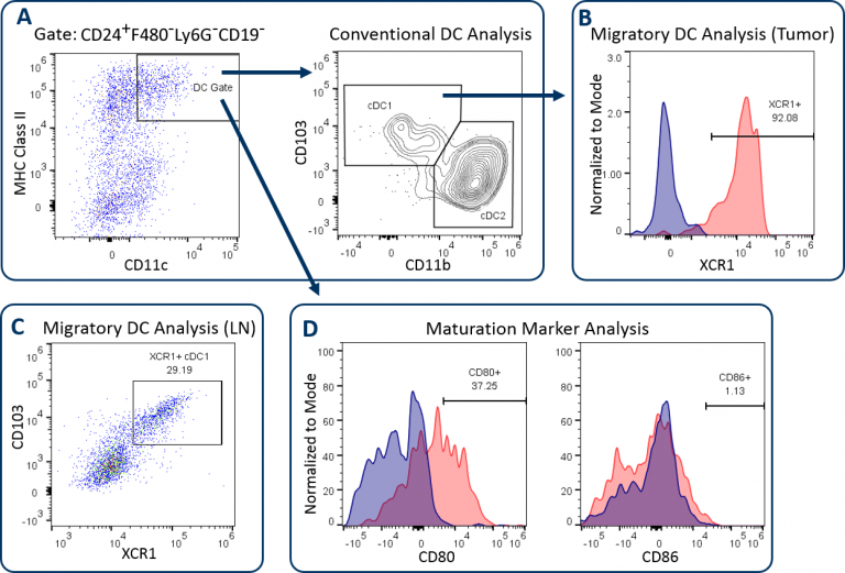
CompDC™面板允许调查人员确定是否亚博全站官网体内治疗影响肿瘤,肿瘤排水淋巴结(LN)或其他宿主隔室中不同DC子集的丰度和激活状态。它是一个12色面板,它结合了一组抗体,当正确检查时,描绘了不同的直流亚群,并向DCS的T细胞激活电位提供了洞察力(表1)。通过细致的门控策略,通过使用排除栅极排除巨噬细胞,粒细胞和B细胞,首先使DC分析促进精确度。该方法最小化由于不相关或自发荧光细胞污染而受到DC分析的风险。然后使用CD24表达有助于描绘常规的DC子集,其通常通过培养基分类为CD11C和MHC II类的高表达水平。这些传统的DC包括CD103+/ cd8.+DC1 subset and the CD11b+DC2子集(图1A)已被证明驱动CD8+和CD4.+T cell anti-tumor responses respectively.[1]XCR1表达已被多组记录为具有交叉呈递活动的DCS的标记,DC介导的CD8激活所需的表型+T细胞。[2]图1B和1C演示了XCR1+B16-F10肿瘤衍生细胞和排水淋巴结的DC分析。XCR1表达式通常与DC1子集相关联,如图所示。最后,测量成熟标志物CD80和CD86的表达,以评估DCS的T细胞共刺激电位。图1D表明,在B16-F10黑色素瘤中的DCS中,这些标志物的表达相对较低,指示肿瘤微环境(TME)在DC上施加的免疫抑制。
表1:CompdC™面板抗体和其实用程序的描述
| 抗体/染料 | 描述 |
|---|---|
| CD45 | 平移免疫细胞标记 |
F4/80, Ly-6G, CD19 |
巨噬细胞,粒细胞和B细胞的排除门 |
| CD11C. | Dendritic cell marker |
| CD24 | Dendritic cell marker |
| CD8 | DC1标记 |
| MHC Class II | 树突状细胞标志物/成熟标记 |
| CD103 | DC1标记 |
| CD11B. | DC2标记 |
| XCR1. | 交叉呈递/迁移树突细胞标记 |
| CD80. | 成熟标记 |
| CD86. | 成熟标记 |
| 活力染料 | 尸体排除 |
| The CompDC™ can be customized to analyze inflammatory DC (InfDC) and plasmacytoid DC subsets by substituting in Ly-6C/CD206 and Siglec-H antibodies respectively. | |
The CompDC™ can also be customized for analysis of two additional DC subsets. This includes inflammatory DCs (infDCs), which is performed by substituting in anti-CD206 and anti-Ly-6C antibodies. InfDCs have been demonstrated to promote anti-tumor activity and can be delineated out of the monocytic myeloid-derived suppressor cell (M-MDSC) gate as CD11c+MHCII.+CD11B.+CD206.+cells (Figure 2A).[3]通过在抗SigleC-H抗体中取代,也可以通过代替血浆谱系DCS(PDC)。虽然TME中的PDC功能尚未完全表征,但许多报告表明这些PDC可能具有损害功能,甚至可以对抗肿瘤反应产生免疫抑制作用。[4]PDCs通常具有低水平的CD11C表达,并且对于SigleC-H是阳性的,如图2b所示。
 Analysis of infDC in B16-F10 tumor-derived cells. (B) Analysis of pDC in CT26 colorectal carcinoma tumor-derived cells.")
要了解有关CompDC™面板的更多信息以及如何将其整合到您的研究中,联系Covance的科学家。
1Gardner, A., & Ruffell, B. (2016). Dendritic cells and cancer immunity.免疫学趋势那37.(12),855-865。
2Wylie,B.,Seppanen,E.,Xiao,K.,Zemek,R.,Zanker,D.,Prato,S.,...与Waithman,J.(2015)。迁移XCR1 + CD103和XCR1 + CD103 +树突细胞的皮肤黑素瘤抗原的交叉呈递。onchoimmunology那4.(8),E1019198。
3.Kuhn,S.,Yang,J.,&Ronchese,F。(2015)。单核细胞衍生的树突细胞对于局部免疫疗法后CD8 + T细胞活化和抗肿瘤反应是必需的。Frontiers in immunology那6.584。
4.Mitchell,D.,Chintala,S.,&Dey,M。(2018)。免疫和癌症中的血浆骨质特性细胞。中国神经杂志杂志。
Note: Studies were performed in accordance with applicable animal welfare regulations in an AAALAC-accredited facility