生物发光成像:肿瘤发展、疾病传播和治疗效果的体内监测
AUTHOR:
Sylvie Kossodo,博士|科学发展副主任
日期:
September 2020
生物发光imaging (BLI) is a non-invasive optical imaging modality designed to visualize and quantify bioluminescent signal in tissues.1BLI是基于酶介导的底物氧化过程中产生的可见光的检测活着o as a molecular reporter. BLI offers a robust, sensitive and high throughput alternative relative to more traditional imaging and biodistribution studies using terminal endpoints, invasive procedures or radiolabeling. Covance was the first CRO to offer BLI in 2003 and in the 17 years since we have amassed considerable experience and know-how in this optical imaging field. In this Technology Spotlight we will present the principles of bioluminescence imaging and highlight advantages this preclinical service offers in cancer detection, monitoring disease progression and体内抗肿瘤疗效评估。
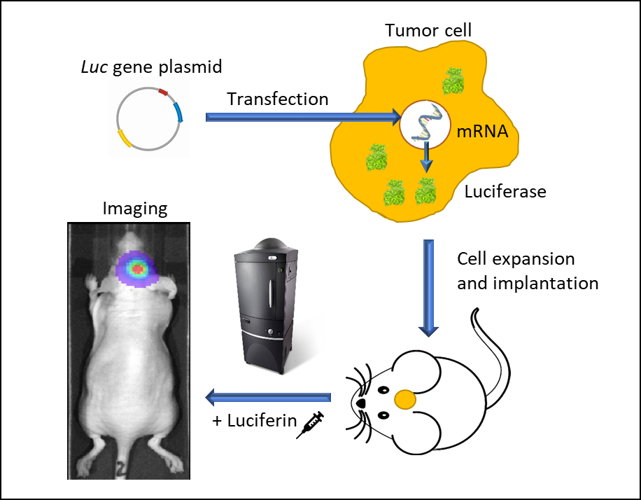
BLI requires cells to express luciferase, a word suitably derived from the Latin路西法-光使者,例如由萤火虫或海三色堇制成。萤火虫Luc.基因在1985年首次克隆2从那时起,绿色是最常用的荧光彩色成像。萤火虫荧光素酶需要注入它的底物D-荧光素,D-荧光素产生562nm的生物发光信号,由置于光密柜中的高量子效率电荷耦合器件相机(CCD)捕获。从工程化肿瘤细胞表达荧光素酶到成像动物的事件序列体内is illustrated in Figure 1. At Covance, we use IVIS® In Vivo Imaging Systems (PerkinElmer, Waltham, MA) which allow high sensitivity and high resolution体内BLI和荧光成像(FLI)在很宽的波长范围内。动物在2%异氟醚气体麻醉下一次最多成像5个。给每只小鼠注射D-荧光素,并在注射后10-15分钟成像。BLI信号在肿瘤周围、特定区域(例如头颅、胸部或腹部)、全身或组织的感兴趣区域(roi)中被量化ex vivo信号表示为每秒光子数,表示从用户定义区域全方位辐射的通量。使用LivingImage4.3.1(PerkinElmer,Waltham,MA)软件分析图像。
原位实体瘤生长监测
Over the past 20 years, the effectiveness and sensitivity of BLI methods and tools have resulted in an ever-broadening catalogue of applications: studies of protein-protein interactions, genetic screening, cell-cycle regulators, parasitic infections, stem cell engraftment tracking, NK cell migration and above all, preclinical oncology. A significant number of studies have shown the ability of BLI imaging to temporally assess tumor development.3This is particularly critical when tumor growth cannot be determined by caliper measurements and relies either on clinical signs such as weight loss or tumor burden assessment after euthanasia of the animals. In the examples shown below animal care and use was conducted according to animal welfare regulations in an AAALAC-accredited facility with IACUC protocol review and approval.
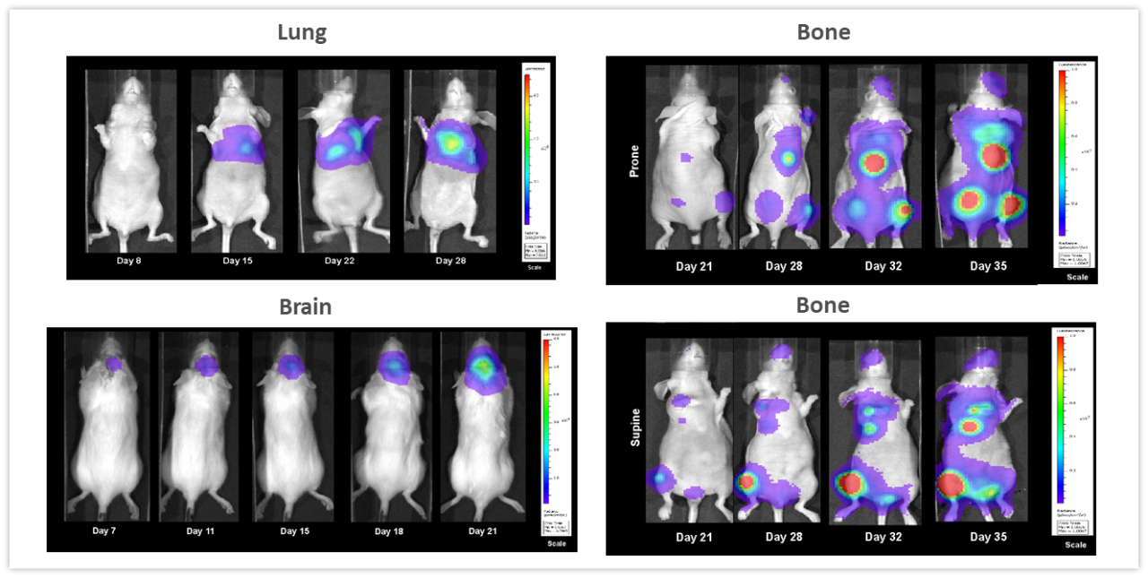
图2显示了强调无创BLI在监测深部组织肿瘤发展中的价值的例子。NCI-H460-Luc2人肺癌异种移植物原位移植(OT)(1x10)5cells/mouse) in the left lung of female nude mice. BLI (Figure 2, top left) show tumors in the thoracic cavity detectable as early as 8 days post tumor cell implantation (not visible in the image due to thresholding to the maximum signal seen on day 28), and tumor burden increasing over the next 2 weeks. Mice experienced a mean percent body weight loss of 18.3% throughout the duration of the study, likely to be disease related (not shown) and were euthanized 28 days after tumor cell implantation. Necropsy revealed significant growth of the primary tumor and substantial development of masses throughout the thoracic cavity. In Figure 2, bottom left, female albino C57BL/6 mice were implanted intracranially with GL261-luc2 syngeneic murine glioma cells (1x10⁶细胞/小鼠)。植入后7天可在大脑中检测到肿瘤。与疾病进展相关的体重减轻在该模型中很常见,可在肿瘤植入后11天检测到,而死亡的中位时间约为21天。人乳腺癌细胞系MDA-MB-231-luc-D3H2LN在心内注射(1x10)后形成骨损伤5(细胞/小鼠)。肿瘤细胞注射后21天,骨损伤清晰可见,在接下来的3周内,肿瘤数量和肿瘤总负荷均增加(图2,右侧,顶部和底部)。小鼠在第32天体重最大下降16.2%。体重减轻主要与模型的攻击性有关(未显示)。从这些研究中得到的信息是,BLI可以成为一个强大的工具,用于实时和非侵入性地研究各种肿瘤类型,而不必仅仅依赖于疾病的临床症状,这些症状可能会或可能不会伴随肿瘤负担的增加。
恶性血液病影像学
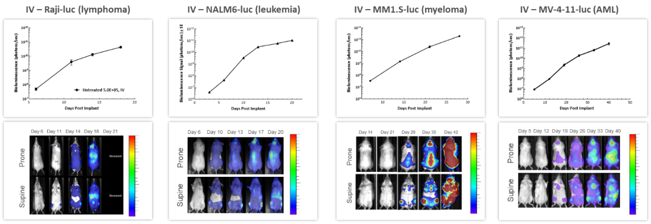
Covance拥有80多个独特的血液恶性肿瘤细胞系,以市场相关的血液恶性肿瘤模型引领行业,特别是过继细胞治疗(ACT),也称为细胞免疫治疗,领域。4在评估血液播散性恶性肿瘤的进展和治疗反应时,BLI是非常有价值的。对静脉注射到NSG小鼠体内的4种不同的人类血液肿瘤细胞进行的纵向研究显示,随着时间的推移,所有模型的肿瘤负担都增加(图3),强调了BLI在反复评估疾病进展和严重程度以及了解肿瘤信号在全身的生物分布方面的价值。
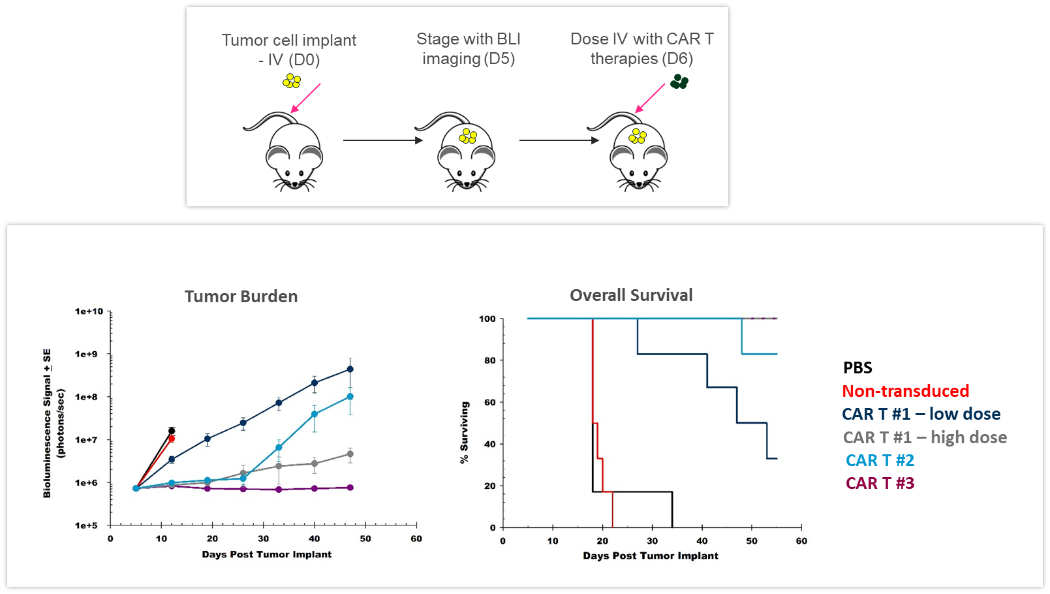
ACT不断发展,为患者提供更好、更个性化的选择。CAR-T细胞是针对肿瘤细胞表达的抗原或标志物的自体或异体T细胞。临床前yaboapp体育官网体内assessment of the efficacy and safety of CAR-T cells is crucial before translating these new therapies into the clinic. The study shown in Figure 4 was designed to test different CAR-T preparations and doses in human Raji-luc B cell lymphoma cells implanted IV in NSG mice. The 3 different CAR-T constructs significantly inhibited tumor burden and prolonged survival, highlighting the value BLI brings to the development of these new cellular immunotherapies.
Monitoring Treatment Efficacy
Radiation
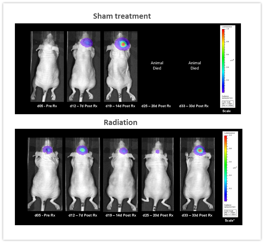
Clearly, one of the key advantages of BLI is the ability to track the efficacy of anti-tumor therapies over time in the same animal, leading to a reduction in the number of animal used. In the example shown in Figure 5, female nude mice were implanted intracranially with NCI-H1975-luc human non-small cell lung carcinoma in order to mimic lung metastasis to the brain. Mice received two courses of fractionated radiation (2Gy; 5 days on, two days off for two cycles), delivered by a RadSource RS-2000 irradiator. The control group was sham irradiated. BLI images were acquired over time and demonstrated that radiation significantly reduced tumor burden (Figure 5, p<0.05 on day 19) and increased life span by 160% (p<0.05, not shown). In this experiment, we were able to quantify treatment response体内, critical when monitoring efficacy as well as designing and refining new therapeutic approaches.
检查点抑制与联合治疗
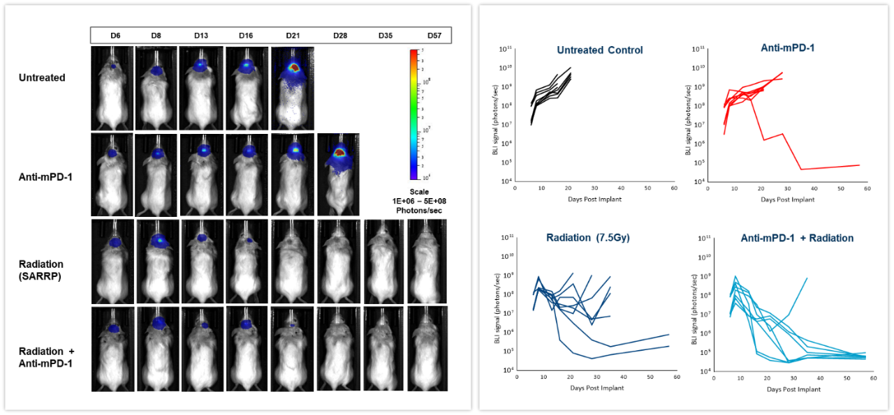
The effect of checkpoint inhibition, alone or in combination with radiation, was tested in murine syngeneic GL261-luc2 glioma cells implanted intracranially in female albino C57BL/6 mice. Mice were left untreated, treated with radiation, delivered by the Small Animal Radiation Research Platform (SARRP; Xstrahl Inc.), at a single 7.5Gy dose, anti-mouse PD-1 (clone RMP1-14, 10 mg/kg) or the combination of both therapies. As shown in Figure 6 (left), BLI signal was already detectable 6 days post-tumor cell implantation and progressed over the next several weeks. Quantification of the resulting bioluminescent signal (right) showed the effect of treatments on tumor burden. Anti-mPD-1 treatment led to 2.6 days tumor growth delay and 1/8 tumor free survivor (TFS) while radiation resulted in 16.6 days tumor growth delay and 2/8 TFS. The combination of radiation and anti-mPD-1 treatment resulted in significant 38.6 days of tumor growth delay and complete remission with 7/8 mice. Traditional methods of gauging anti-tumor response in malignancies located deep within the body are based on terminal and often, time consuming read-outs. As shown in these examples, the integration of体内BLI成像使我们能够从肿瘤治疗的“黑匣子”转变为对个体反应的实时评估,从而提供更快速调整和完善治疗的灵活性。
多模态
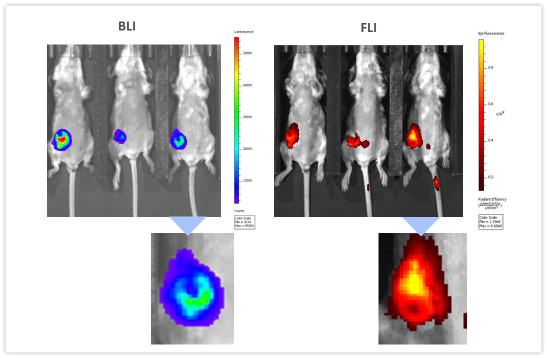
BLI可以与其他成像方式结合,以询问不同的生物途径。为了说明这个应用,我们使用了同基因4T1-luc2-1A4mouse breast adenocarcinoma cells injected OT into the mammary fat pad of immunologically competent BALB/c mice. When tumors reached ~300 mm3, a near-infrared imaging agent, ProSenseTM750注射IV以检测与侵袭性乳腺癌生长相关的组织蛋白酶活性。prosTM750是一个光无声的代理,它变成了流感orescent upon cleavage with cathepsins. As shown in Figure 7, BLI and FLI tumor signals were readily detectable in tumors. However, BLI and FLI signals were not perfectly superimposable which is expected given that luciferase is produced by all living tumor cells while the fluorescent signal is confined not only to tumor cells but also other tumor-associated cells, mainly macrophages, responsible for the cleavage and uptake of the cathepsin ProSenseTM750探头。因此,多模态可以用来审问不同的生物学体内,同时。
概要
Mouse models of deep-tissue cancer and cancer metastasis rely predominantly on terminal assessments like tissue weight, nodule counts, and/or histologic analysis for the assessment of tumor burden. Since its introduction well over 20 years ago, BLI has become an invaluable technique to track the growth dynamics of bioluminescently-tagged cancer cell lines over time and in response to various therapeutic approaches体内,最大限度地减少研究中的动物数量,因为相同的受试者可以随时间成像,而无需进行最终评估。BLI在许多科学领域都做出了重大贡献,除了为使用已有的侵入性或依赖放射性同位素的非侵入性成像方式提供了一种替代方法。
联系科文斯的科学家to request the full data set or to learn more about our BLI service and how it can be applied to your preclinical research.
1Contag CH, Spilman SD, Contag PR, Oshiro M, Eames B, Dennery P, Stevenson DK, Benaron DA. Visualizing gene expression in living mammals using a bioluminescent reporter. Photochem Photobiol 1997; 66: 523–531.
2de Wet JR,Wood KV,Helinski DR,DeLuca M.萤火虫荧光素酶cDNA的克隆及其在小鼠体内的表达Escherichia coli. 过程。纳特尔。阿卡德。科学院。U、 S.A.1985;827870–7873。
3Jenkins DE, Oei Y, Hornig YS, Yu SF, Dusich J, Purchio T, Contag PR. Bioluminescent imaging (BLI) to improve and refine traditional murine models of tumor growth and metastasis. Clin Exp Metastasis, 2003; 20(8):733-744.
4https://www.cancerresearch.org/immunotherapy/treatment-types/adoptive-cell-therapy
